Archive of

Understanding Depression: Its Impact on the Brain and how to deal with it

Depression is a pervasive mental health condition that affects millions of people worldwide. While it often manifests as emotional distress, its roots lie deep within the brain's intricate network of neurons and neurotransmitters. In this article, we'll explore how depression affects the brain and delve into non-pharmacological interventions that can help individuals manage and overcome this debilitating condition.

The Brain and Depression

To understand how depression affects the brain, it's crucial to recognize that it is not solely a "mind" issue but a complex interplay between biology, genetics, environment, and psychology. Several key brain regions and neurotransmitters play significant roles in depression:

  1. The Prefrontal Cortex: This area is responsible for executive functions like decision-making and problem-solving. In people with depression, it often shows reduced activity, leading to difficulties in concentrating and making choices.

  2. The Amygdala: The amygdala plays a central role in processing emotions, particularly negative ones. It tends to be hyperactive in individuals with depression, leading to heightened sensitivity to stressors and increased feelings of sadness.

  3. Hippocampus: The hippocampus is involved in memory and learning. In those with depression, it often shrinks in size, which may contribute to memory problems and difficulties in processing information.

  4. Neurotransmitters: Brain chemicals like serotonin, dopamine, and norepinephrine are essential for mood regulation. Depression is often associated with imbalances in these neurotransmitters, affecting mood, sleep, and appetite.

Non-Pharmacological Interventions for Depression

While medications can be effective in treating depression, non-pharmacological interventions provide valuable alternatives, especially for individuals who prefer a drug-free approach or want to complement their medication regimen. Here are some evidence-based non-pharmacological interventions:

  1. Psychotherapy: Cognitive-behavioral therapy (CBT), interpersonal therapy (IPT), and mindfulness-based cognitive therapy (MBCT) are proven psychotherapeutic approaches for depression. They help individuals identify and change negative thought patterns and develop healthy coping strategies.

  2. Exercise: Physical activity releases endorphins, the body's natural mood elevators. Regular exercise not only improves mood but also reduces stress and anxiety. Even simple activities like walking or yoga can be beneficial.

  3. Nutrition: A balanced diet rich in nutrients can support overall brain health. Omega-3 fatty acids, found in fish and flaxseeds, have been linked to improved mood. Avoiding excessive sugar and processed foods can also help stabilize mood.

  4. Sleep Hygiene: Depression often disrupts sleep patterns, and poor sleep can exacerbate depressive symptoms. Establishing a regular sleep schedule and creating a calming bedtime routine can improve sleep quality.

  5. Social Support: Isolation can worsen depression. Engaging in social activities and maintaining strong social connections can provide emotional support and reduce feelings of loneliness.

  6. Mindfulness and Meditation: Mindfulness practices can help individuals become more aware of their thoughts and emotions without judgment. Mindfulness-based stress reduction (MBSR) and mindfulness-based cognitive therapy (MBCT) have shown promise in reducing depressive symptoms.

  7. Stress Management: Learning stress reduction techniques like deep breathing, progressive muscle relaxation, or biofeedback can help individuals manage the physiological and emotional aspects of stress.

  8. Art and Music Therapy: Creative outlets such as art and music therapy can provide a non-verbal means of expressing and processing emotions, reducing the burden of verbal communication.

Conclusion

Depression is a complex mental health condition that impacts not only emotions but also the brain's physical structures and chemical processes. While medications can be effective, non-pharmacological interventions offer valuable options for individuals seeking drug-free approaches or supplementary strategies. By understanding how depression affects the brain and exploring these non-pharmacological interventions, individuals can better manage and eventually overcome the challenges posed by this condition. Remember that seeking professional help is crucial, as a combination of treatments tailored to individual needs often yields the best results in managing depression.
The Memory Game: How Synaptic Potentiation Keeps Your Brain Sharp

Hey there, memory enthusiasts! Ever wondered how your brain manages to remember your best friend's birthday, the lyrics to your favorite song, or even where you left your keys? Well, it's all thanks to a fascinating process called synaptic potentiation. Don't worry, we're going to break it down in a way that's as easy to digest as your favorite snack.

What's Synaptic Potentiation Anyway?

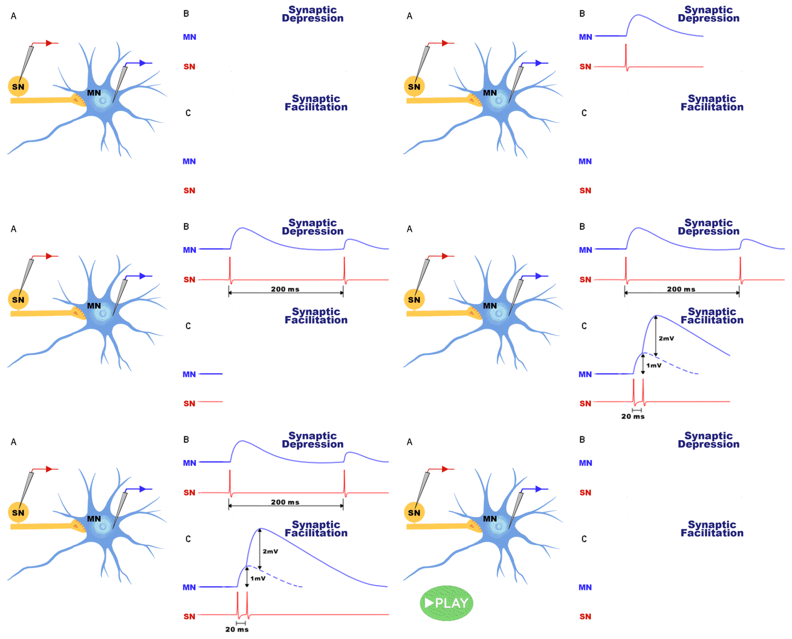
Alright, let's dive in. Synaptic potentiation is like the secret sauce of memory retention. Imagine your brain is a big, complex puzzle, and the puzzle pieces are your neurons. These neurons communicate with each other at junctions called synapses. Synaptic potentiation is all about making these synapses stronger and more efficient, like supercharging your brain's connections.

The Learning Process

So, here's how it works. When you learn something new, like riding a bike or learning a new dance move, your brain forms new connections between neurons. These connections are a bit like electrical circuits that transmit information.

Initially, these connections are like dirt paths in the woods—weak and hard to follow. But as you keep practicing that new skill or cramming for a test, something incredible happens. Your brain strengthens these connections through a process called long-term potentiation (LTP). It's like turning those dirt paths into superhighways!

The Role of Neurotransmitters

Now, let's talk about neurotransmitters. These are like little messengers in your brain that help neurons communicate. One neurotransmitter that plays a crucial role in LTP is glutamate. When you're learning and experiencing something new, your brain releases more glutamate, which helps in strengthening the synaptic connections.

Imagine glutamate as the construction workers who come in and reinforce your superhighways, making them even wider and more efficient.

The Importance of Repetition

Repetition is key to memory retention. The more you practice or revisit something, the stronger those synaptic connections become. It's like constantly renovating and expanding your superhighways to accommodate more traffic.

Think about it like this: the first time you meet someone, you might forget their name pretty quickly. But if you keep meeting them and saying their name, your brain says, "Hey, this person is important!" and reinforces the memory.

Sleep and Memory Consolidation

Here's a fun fact: your brain doesn't just strengthen these connections while you're awake and learning. It also works its magic during your sleep. That's right, good old-fashioned snooze time is when your brain consolidates memories and enhances those newly formed pathways.

So, next time someone tells you to get a good night's sleep before a big test, you can thank synaptic potentiation for helping you ace it!

Wrap-Up

In a nutshell, synaptic potentiation is the brain's way of saying, "I've got your back when it comes to remembering stuff." It's like upgrading your mental toolkit to keep your memories fresh and accessible.

So, whether you're learning a new language, mastering a musical instrument, or simply trying to remember where you left your phone, give synaptic potentiation a big thumbs up. It's the unsung hero behind your incredible memory!

Unlocking the Potential of Academic Search Engines for Effective Research – feat Related Engine

In the vast landscape of academic and scholarly research, the ability to navigate through the ocean of information is crucial for researchers and students alike. Academic search engines play a pivotal role in simplifying this process, offering access to a wealth of valuable resources. In this article, we'll explore how to master academic search engines for research and introduce you to Related Engine relengine.com as one of the tools for finding and navigating through academic and scholarly research results. The Power of Academic Search Engines

Academic search engines have revolutionized the way researchers and students access scholarly content. These platforms aggregate a wide range of academic sources, including journals, articles, theses, conference papers, and more. They provide a convenient, user-friendly interface to help users sift through the vast sea of information and pinpoint the most relevant resources for their research. Now, How to Master Academic Search Engines? To make the most of academic search engines, you need a strategic approach. Here are some tips to help you master the art of research using these platforms:

  1. Refine Your Search Queries Crafting effective search queries is the first step in mastering academic search engines. Start with a clear research question or topic and use relevant keywords to refine your search. Boolean operators like "AND," "OR," and "NOT" can help you narrow or broaden your search results.
  2. Utilize Advanced Search Features Most academic search engines offer advanced search features that allow you to filter results by publication date, author, journal, and more. Take advantage of these features to pinpoint the most relevant and up-to-date sources for your research.
  3. Use Citation Searches Citation searches are a valuable tool for finding related research. If you come across a seminal paper or article in your field, use the citation feature to explore other works that have referenced it. This can lead you to additional relevant sources, you can track papers citations using either our PubMed search engine database, or a diverse group of advanced academic search engines databases. 1- https://search.relengine.com/pm 2- https://search.relengine.com/ai

  4. Explore Related Articles Many academic search engines provide a list of related articles or papers based on your search query. These can be a goldmine of additional resources that you might have missed otherwise. We also introduce different ways to track related article, with graphical representation through all of our search resources check them out https://relengine.com

  5. Create Alerts Stay up-to-date with the latest research in your field by setting up alerts. Many academic search engines offer alert features that notify you when new papers matching your criteria are published. Related Engine. In your journey to master academic search engines, Related Engine is a valuable ally. This academic search engine helper tool is designed with the researcher in mind with a user friendly interface, extensive database of academic sources, and diverse search capabilities. Here's why it's worth considering:
  6. Comprehensive Database Related Engine searches in an extensive database of academic and scholarly research sources, covering a wide range of disciplines. Whether you're studying the sciences, humanities, social sciences, or any other field, you'll find relevant content here.
  7. User-Friendly Interface Navigating through Related Engine results is a breeze. The intuitive interface ensures that you can quickly locate and access the resources you need without any steep learning curve.
  8. Citation and Related Articles With features like citation searches and related articles, Related Engine relengine.com makes it easy to discover connections between different research papers and explore additional sources that can enhance your research.

We’d be happy to give our tool a try, and happy exploring.

Related Engine Search Tools

مرض التصلب المتعدد "MS" : التعامل والعلاجات المتاحة

يعتبر التصلب المتعدد مرضًا محيرًا للغاية، حيث يختلف تأثيره وتقدمه من شخص لآخر. يعاني البعض من أعراض خفيفة تتطور ببطء، في حين يواجه البعض الآخر أعراضًا حادة وتقدم سريع. بالرغم من ذلك، هناك عدد من الإجراءات التي يمكن اتخاذها للتعامل مع هذا المرض وتحسين الجودة الحياتية للمصابين به.

التصلب المتعدد هو مرض مزمن يؤثر على الجهاز العصبي المركزي، ويعد واحدًا من أكثر الأمراض التي تؤثر على الجهاز العصبي شيوعًا. يُعرف التصلب اللويحي بأنه مرض يهاجم الغلاف الواقي للأعصاب (الميالين) ويسبب تلفًا في الأعصاب، مما يؤدي إلى تشوهات في نقل الإشارات العصبية ويمكن أن يتسبب في مجموعة متنوعة من الأعراض.

كيفية تشخيص التصلب المتعدد؟

تشخيص التصلب المتعدد يتضمن عدة اختبارات وإجراءات. يبدأ الأمر بتقييم الأعراض والتاريخ الصحي للمريض. يتبع ذلك إجراء اختبارات التصوير بالرنين المغناطيسي للدماغ والحبل الشوكي، والتي تساعد في رصد التلف في الأعصاب. أيضًا قد تُجرى اختبارات أخرى مثل التصوير بالأشعة السينية وفحص الدم لاستبعاد أمراض أخرى.

التعامل مع التصلب المتعدد:

الدعم النفسي والاجتماعي: يعد الدعم النفسي والاجتماعي من أهم العوامل في مساعدة المرضى على التعامل مع التصلب المتعدد. يمكن أن يكون التحدث مع أشخاص آخرين مصابين بنفس المرض أو البحث عن دعم من خلال مجموعات دعم مفيدًا جدًا.

العلاج الطبيعي والعلاج الوظيفي: يمكن أن يساعد العلاج الطبيعي والعلاج الوظيفي في تحسين القوة العضلية والتوازن، وتعزيز القدرة على التنقل وأداء الأنشطة اليومية بشكل أفضل.

الأدوية: يتم استخدام الأدوية المضادة للالتهاب والأدوية المثبطة للمناعة لإدارة وتقليل التضخم والتلف في الجهاز العصبي المركزي. تختلف الأدوية المستخدمة حسب حالة المريض وتقدم المرض.

العلاجات المتاحة:

بالإضافة إلى العلاجات المذكورة أعلاه، هناك أبحاث مستمرة وتطورات في مجال علاج التصلب المتعدد. يعمل العلماء والباحثون جاهدين لفهم أفضل طبيعة المرض واكتشاف علاجات جديدة.

مقترحات لأسلوب حياة يومي لمريض التصلب المتعدد وأهله:

تنظيم الوقت: تنظيم اليوم وجدولته بعناية يمكن أن يساعد في تقليل التوتر وزيادة الإنتاجية.

ممارسة الرياضة: مع استشارة الطبيب، يمكن لمرضى التصلب المتعدد الاستفادة من ممارسة الرياضة بانتظام للمحافظة على لياقتهم البدنية.

التغذية السليمة: يجب أن يكون النظام الغذائي غنيًا بالفيتامينات والمعادن والألياف لدعم الصحة العامة.

الاسترخاء والتأمل: تقنيات التأمل والاسترخاء يمكن أن تساعد في تقليل التوتر وتحسين العافية النفسية.

البقاء مستندين إلى المعلومات: تعلم المزيد عن المرض والتوجيهات الطبية يمكن أن يساعد في فهم أفضل للتصلب المتعدد وكيفية التعامل معه.

في النهاية، التصلب المتعدد قد يكون تحديًا كبيرًا، ولكن مع العلاج الصحيح وتبني أسلوب حياة صحي، يمكن للمصابين به أن يعيشوا حياة مستقرة ومرضية.

Habits and the Brain: When Dopamine and Serotonin Join the Fun!

Hey there, fellow habit aficionado! Habits, huh? They're like those quirky little routines that make us who we are, from that daily caffeine fix to the ritual dance you do before bedtime. But did you ever stop to wonder what's happening inside that noggin of yours when you're deep into your habits? Well, that's where dopamine and serotonin step into the limelight – they're the brain's dynamic duo that can make you feel all sorts of things, including those bursts of happiness and satisfaction that keep you hooked. In this blog post, let's get cozy with these brain chemicals and see how they spice up our habits. Oh, and don't worry, I've got some jokes to keep things light and fun!

The Brain's Stand-Up Comedy Show: Before we dive into the nitty-gritty, let's tickle those funny bones with some habit-related humor:

Why did the neuron refuse to hang out with dopamine anymore? Because it just couldn't resist its addictive charm!

How does a serotonin molecule like to unwind on weekends? Kicking back on the beach, riding the waves of relaxation, of course!

What did one habit say to the other habit? "We should stop meeting like this; we're getting way too predictable!"

The Science of Habit Formation: Now, let's get a bit serious (but not too much, because who wants that?) and talk about how dopamine and serotonin shape our habits.

Dopamine: The Motivator

Dopamine is often called the "feel-good" neurotransmitter. It's like your brain's way of saying, "Hey, you're doing something awesome!" When you achieve a goal or find pleasure in something, dopamine levels skyrocket, creating this rush of motivation and satisfaction. This is why you get that little pat on the back feeling when you complete a task or savor a delicious treat.

Here's a joke for your dose of dopamine: Why did the procrastinator's brain flood with dopamine? Because it knew how to live on the edge of deadlines!

Serotonin: The Mood Balancer

Serotonin, on the other hand, is like the zen master of your brain. It's all about keeping your emotions in check, managing anxiety, and ensuring you have those moments of calm. Low serotonin levels can lead to feelings of sadness and stress, while higher levels bring on that warm, fuzzy feeling of well-being. Serotonin is often the reason you feel so content after a good workout or a relaxing evening with friends.

And now, a serotonin-inspired joke: Why did the serotonin molecule start meditating? To find its inner peace, of course!

The Habit Loop:

These two neurotransmitters, in their charming, chemically charged way, work together to create habits. You've got your cue, your routine, and your reward. When you dive into a habit, dopamine does a little happy dance in anticipation of the reward, pushing you to repeat the routine. As time goes on, your brain wires itself to crave that habit, and voilà, it becomes an integral part of your daily life.

So, whether it's hitting the gym, nibbling on your favorite chocolate, or indulging in a Netflix marathon, your brain's the real MVP, orchestrating the release of dopamine and serotonin to keep you coming back for more.

Conclusion: So, there you have it, the delightful duo in your brain – dopamine and serotonin – working their magic to make habits stick. It's all about motivation, mood, and a dash of chemistry that keeps us going. Next time you catch yourself in a habit loop, just remember these two brainy buddies and maybe toss in a joke or two to keep things fun. After all, life's a habit worth enjoying, one laugh at a time!

Unlocking the Secrets of Your Brain's Superpower: Memory!

Hey there, 🧠💪

Ever wondered how your brain manages to remember things like where you left your keys, your favorite childhood ice cream flavor, or even the lyrics to that catchy song you heard once? Well, you're not alone! Let's dive into the fascinating world of memory in everyday terms.

The Brain's Marvelous Memory:

Imagine your brain as a superhero, and memory is its special power. It's like a vault storing all the information you've ever come across, and it's constantly working behind the scenes, helping you navigate life.

Short-Term vs. Long-Term Memory:

Your brain has two memory modes: short-term and long-term. Short-term memory is like your brain's sticky note—it holds information for a brief time, like a phone number you jot down temporarily. Long-term memory is where the magic happens. It's like a library filled with your life's stories, ready to be accessed whenever needed.

How Memories Form:

Memories are created when your brain forms connections between different neurons, which are like tiny messengers in your brain. These connections become stronger with repetition and emotion. So, that's why you remember your childhood best friend's name, even if you haven't seen them in years!

Tips to Boost Your Memory:

Stay Curious: Engage your brain with new experiences and knowledge. Learning keeps your memory sharp.

Get Quality Sleep: Sleep helps consolidate memories. So, make sure you catch those Z's!

Stay Active: Regular exercise boosts brain health and memory.

Healthy Eating: Nutrient-rich foods are brain fuel. Think fruits, veggies, and Omega-3 fatty acids.

Mindfulness: Reduce stress through meditation or mindfulness exercises. Stress can be memory's enemy.

Remember (pun intended) – Your Brain is Unique:

No two brains are the same. What you remember might be different from what someone else does. Embrace your brain's uniqueness, and don't be too hard on yourself.

So, there you have it, a quick peek into the world of your brain's memory superpower! Keep feeding your brain with experiences, knowledge, and love, and watch it work its magic. 🌟

Got any memory-related questions? Share them below! Let's chat about the amazing world inside our heads! 🤓🧠 #BrainPower #MemoryMagic #CuriousMinds

Navigating the Brain's Blueprint: A Guide to Using the Human Connectome Project

The Human Connectome Project (HCP) is like a treasure map for understanding the inner workings of our brains. It provides a detailed roadmap of the brain's structural and functional connections, shedding light on the complex web that orchestrates our thoughts, emotions, and actions. In this blog post, we'll explore how to use the HCP to delve into this fascinating realm of neuroscience. Get ready to embark on a journey through the neural pathways of the mind!

Step 1: Accessing the HCP Data

The first step on our journey is to access the rich dataset provided by the HCP. Follow these steps:

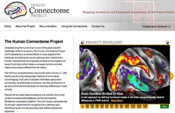
Visit the HCP Website: Start by going to the Human Connectome Project website.

Register for Access: To access the data, you'll need to create an account and agree to the terms of use. This ensures responsible and ethical use of the data.

HCP Website

Step 2: Understanding the HCP Data

Before diving into the data, it's essential to understand what you'll find. The HCP offers two main types of connectome data: structural and functional.

Structural Connectome: This data represents the physical wiring of the brain, akin to mapping the roads and highways of a city. It's obtained using Diffusion MRI (dMRI) technology.

Structural Connectome

Functional Connectome: Think of this as tracking the flow of traffic in the city. Functional connectome data comes from Resting-State Functional MRI (rs-fMRI) scans and task-based functional imaging.

Functional Connectome

Step 3: Navigating the Connectome Data

Now, let's explore how to navigate and visualize the connectome data:

Connectome Workbench: The HCP provides a powerful tool called the Connectome Workbench. Download and install it on your computer.

Connectome Workbench

Loading Data: Use the Connectome Workbench to load your desired connectome dataset, whether structural or functional.

Loading Data

Step 4: Exploring the Connectome

With the data loaded, you're ready to start exploring the connectome:

Visualizing Networks: You can visualize brain networks and connections in 3D using the Connectome Workbench. This helps you understand how different brain regions communicate.

Visualizing Networks

Analyzing Connectivity: Dive into the data to analyze connectivity patterns. Look for connections that might be associated with specific brain functions or disorders.

Analyzing Connectivity

Step 5: Applying the Knowledge

The insights gained from the HCP data can be applied in various fields, from neuroscience research to medical diagnostics and treatment. Researchers can use this data to study brain disorders, understand individual variability, and explore brain-behavior relationships.

Happy exploring!

Navigating the Mysteries of the Mind: The Human Connectome Project

NIH

The human brain is often regarded as the most complex and intricate organ in the human body. Its billions of neurons and trillions of synapses work in harmony to create our thoughts, emotions, and behaviors. Understanding the intricate web of connections within the brain is a monumental challenge that has fascinated scientists for centuries. In the quest to unravel the mysteries of the mind, the Human Connectome Project has emerged as a groundbreaking initiative, shedding light on the intricate network that underpins our cognition, emotions, and behaviors.

I. What is the Human Connectome Project?

The Human Connectome Project (HCP) is a collaborative research effort that aims to map and comprehensively understand the connectivity of the human brain. Launched in 2010, this ambitious endeavor represents a fusion of neuroscience, advanced imaging technology, and data science. It seeks to provide a detailed blueprint of the brain's structural and functional connectivity, akin to mapping the highways and byways of a vast, interconnected city.

II. Mapping the Structural Connectome

Diffusion Magnetic Resonance Imaging (dMRI)

The structural connectome focuses on mapping the physical pathways of neural connections within the brain. dMRI is a pivotal technology employed in the HCP, allowing scientists to visualize the white matter tracts that connect different brain regions. By tracing the movement of water molecules along axons, dMRI generates images that reveal the structural connections between regions, providing valuable insights into the brain's wiring. High-resolution Brain Scans

The HCP employs high-resolution neuroimaging techniques to capture detailed anatomical information. Combining data from multiple imaging modalities, such as T1-weighted and T2-weighted MRI, enables researchers to create accurate structural maps of the brain. III. Uncovering the Functional Connectome

Resting-State Functional Magnetic Resonance Imaging (rs-fMRI)

The functional connectome explores how different brain regions communicate and synchronize their activities. rs-fMRI measures the blood flow and oxygenation in the brain, allowing researchers to identify patterns of activity across various regions. This technique reveals functional connections, shedding light on the brain's dynamic interactions during both rest and task-related activities. Task-Based Functional Imaging

In addition to resting-state data, the HCP collects data from participants engaged in various cognitive tasks. These task-based functional imaging experiments help researchers understand how different brain networks collaborate to perform specific functions, such as memory, attention, or language processing. IV. The Challenge of Big Data

The HCP generates vast amounts of complex data, necessitating cutting-edge computational methods and data analysis tools. The project's data-sharing philosophy encourages collaboration and data accessibility, enabling researchers worldwide to explore and analyze the rich dataset.

V. Insights and Discoveries

Over the years, the Human Connectome Project has yielded numerous insights into the organization and function of the human brain:

Individual Variability: The HCP has highlighted the significant variability in brain connectivity among individuals, challenging traditional notions of a "typical" brain.

Brain Disorders: Mapping the connectome has also provided valuable insights into the neurological basis of various brain disorders, including autism, schizophrenia, and Alzheimer's disease.

Brain-Behavior Relationships: Researchers have identified strong associations between the brain's connectivity patterns and individual differences in behavior, cognition, and personality traits.

Conclusion

The Human Connectome Project represents a pioneering effort to decode the intricate neural pathways that shape our thoughts, emotions, and behaviors. As technology and data analysis techniques continue to advance, the project holds the promise of unlocking even more profound insights into the human brain. Ultimately, a comprehensive understanding of the connectome may lead to breakthroughs in diagnosing and treating neurological and psychiatric disorders, paving the way for a brighter future in neuroscience and healthcare.

Unmasking the Power of Fear: Investigating Fear Conditioning in Neuroscience

Fear is an integral part of the human experience, a primal emotion that has evolved over millennia to help us survive in a dangerous world. Understanding the neural mechanisms behind fear is not only fascinating but also crucial for unraveling the mysteries of the human brain. One of the fundamental ways neuroscientists study fear is through a process known as fear conditioning. In this blog post, we will delve into what fear conditioning is, how it is investigated in neuroscience, and why it holds the key to understanding our complex emotional responses.

The Basics of Fear Conditioning

Fear conditioning is a form of associative learning in which an organism learns to associate a neutral stimulus (such as a sound or a visual cue) with an aversive or fearful experience. The classic example is Pavlov's experiments with dogs, in which he paired the sound of a bell with the presentation of food. Over time, the dogs began to salivate at the mere sound of the bell, even in the absence of food. In fear conditioning, the process is similar, but instead of salivation, the organism develops a fear response to the previously neutral stimulus.

Fear conditioning is a valuable tool for studying the neural basis of fear and has broad applications in both animal and human research. By investigating the neural pathways and mechanisms involved in fear conditioning, scientists can gain insights into various psychiatric disorders such as anxiety disorders and post-traumatic stress disorder (PTSD).

Investigating Fear Conditioning in Neuroscience

Animal Studies Animal studies, primarily conducted in rodents like mice and rats, have been instrumental in unraveling the neural circuits underlying fear conditioning. Here's a simplified breakdown of the process:

a. Training Phase: Researchers expose the animals to a neutral stimulus, followed by an aversive stimulus, such as a mild electric shock. The animals learn to associate the two stimuli.

b. Testing Phase: After the animals have learned the association, scientists can investigate their fear responses. This can be done through behavioral tests, like measuring freezing behavior (a common fear response in rodents), or through neuroimaging techniques to monitor brain activity.

c. Neural Mapping: Cutting-edge techniques, such as optogenetics and chemogenetics, allow researchers to manipulate specific neural circuits to observe their roles in fear conditioning. This involves controlling the activity of neurons in real-time to understand their contributions to fear learning and expression.

Human Studies Studying fear conditioning in humans involves a different approach due to ethical constraints. However, researchers have developed paradigms that simulate fear conditioning without causing harm. One common method is the use of conditioned fear responses to visual or auditory cues.

a. Experimental Setup: In a controlled laboratory setting, participants are exposed to a neutral stimulus (like a colored shape) paired with an aversive stimulus (such as a loud, unpleasant sound). Over time, participants develop fear responses to the neutral stimulus.

b. Neuroimaging: Functional Magnetic Resonance Imaging (fMRI) and electroencephalography (EEG) are commonly used to monitor brain activity during fear conditioning experiments. Researchers can identify which brain regions are involved and how they communicate.

c. Clinical Applications: Understanding fear conditioning in humans has important implications for treating conditions like PTSD and phobias. Exposure therapy, a common treatment for such conditions, is based on principles of fear extinction, which is closely related to fear conditioning.

Conclusion

Fear conditioning is a powerful tool in neuroscience that helps researchers uncover the intricate neural mechanisms underlying our fear responses. Through animal and human studies, scientists have made significant strides in understanding how fear is acquired and processed in the brain. This knowledge not only enhances our understanding of basic neuroscience but also has far-reaching implications for the treatment of anxiety disorders and trauma-related conditions. As technology continues to advance, fear conditioning research promises even greater insights into the depths of our emotional experiences.